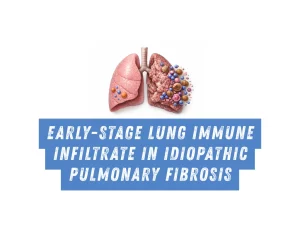
Different Immune Infiltrate in Idiopathic Pulmonary Fibrosis Patients With Stable and Progressive Disease

Objectives and Research Methods in Idiopathic Pulmonary Fibrosis
Disease progression is highly variable in patients with Idiopathic Pulmonary Fibrosis (IPF) and there are no clinical or biological markers to predict it. In our previous works we described two subgroups of end stage IPF patients with different lung immune infiltrate.
Objectives:To compare the lung immune infiltrate in early IPF stages and associate it with the disease progression.
Methods: Diagnostic biopsy samples of 8 patients with IPF were stained with 27 antibodies conjugated with metals and acquired using Imagine Mass Cytometry (IMC) with the Hyperion (Fluidigm).
The panel comprehensively covered more than 20 immune cell types and subpopulations. Patients were clinically followed up for 1 to 10.
Disease progression was defined as above annual Forced Vital Capacity (FVC) decline >= 10%, Diffusing Capacity for Carbon Monoxide (DLCO) decline >= 15% and/or death within the first-year of follow-up.
Results Linking Immune Infiltrate and Idiopathic Pulmonary Fibrosis Progression
IPF progressors presented a lung immune infiltrate enriched in B cells while patients with stable disease had more T cells. NK cells were present in similar proportions but the progressors exhibited an altered NK subpopulations lacking granzyme B.
IPF progressors showed increased expression of adaptive immune cell activation markers such as HLA-DR.
Further analyses are ongoing to study the cell-cell interactions and differences regarding the histopathological region.
Clinical Implications for Idiopathic Pulmonary Fibrosis
Conclusions: Differences in the early-stage IPF lung immune infiltrate associate with disease progression suggesting a key role of specific immune cell types in the pathobiology of the disease.
Authors
Cruz Santa Cruz, T., Hernandez-Gonzalez, F., Martinez, D., Casas-Recasens, S., Agusti, A., Sellares Torres, J., Faner, M.R.
Read more details at
Fecha de publicación
Published Print: 2025-05
Categorías asociadas al artículo
Noticias relacionadas
Cellular Senescence Markers and Matrix Metalloproteinase-7 Are Co-expressed in Fibrosing Interstitial Lung Diseases
Study highlights the role of cellular senescence markers (p16, p21) and MMP-7 co-expression in fibrosing interstitial lung diseases.
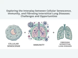
Exploring the Interplay between Cellular Senescence, Immunity, and Fibrosing Interstitial Lung Diseases: Challenges and Opportunities
Discover how cellular senescence, marked by antagonistic pleiotropy, and immune dysregulation play a detrimental role in the pathogenesis of fibrosing interstitial lung diseases (f-ILDs) and pulmonary fibrosis.
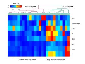
Lung immune signatures define two groups of end-stage IPF patients
To investigate it, we calculated immune signatures with Gene Set Variation Analysis (GSVA) and applied them to the lung transcriptome followed by unbiased cluster analysis of GSVA immune-enrichment scores, in 109 IPF patients from the Lung Tissue Research Consortium (LTRC)
Imagen desarrollada con Canva